Efficacy of Sodium Ascorbyl Phosphate in the Removal of Dental Caries in Canines: A Preliminary Clinical Trial
Ⓒ The Korean Environmental Sciences Society. All rights reserved.
This is an Open-Access article distributed under the terms of the Creative Commons Attribution Non-Commercial License (http://creativecommons.org/licenses/by-nc/3.0) which permits unrestricted non-commercial use, distribution, and reproduction in any medium, provided the original work is properly cited.
Abstract
Dental caries and tartar can lead to infections and gum disease. Appropriate techniques are required to remove dental caries and tartar from companion canines to improve their oral healths. The present study investigated the efficacy of sodium ascorbyl phosphate (vitamin C derivative, STAY-C®50) for dental plagues removal in canines, as a preliminary clinical trial. The canines were randomly divided into two groups from three each: control group (CG, no treatment) and treatment group (TG, Treatment with STAY-C®50). In the TG, sodium ascorbyl phosphate (700 mg) was dissolved in water and applied to the gums near the tooth roots using a syringe in the morning and evening. Photographs were taken using QScan Plus equipment at 0, 2, 4, 6, 10, 12, 14, 16, and 20 weeks to access the efficacy of sodium ascorbyl phosphate in removing dental caries. The treatment group showed reduced dental plaque areas in the incisors and canines, and in the incisors alone, compared to the control group. However, the differences between the two groups were not significant (p>0.05). No significant differences in the ratios of clear dental area to dental plaque area of the incisors were observed at 0–6 weeks; however, significant differences were observed after 10 weeks (p<0.05). Changes in the clear dental and dental plaque areas were significantly greater in the treatment group than in the control group. The findings of this study highlight the positive effects of sodium ascorbyl phosphate on dental plagues removal in canines.
Keywords:
Canine, Clear dental area, Dental plaque area, Incisors, Sodium ascorbyl phosphate1. Introduction
In modern society, owning a companion animal is a common phenomenon that improves the quality of life of lonely people who lack close human relationships (Kim and Youn, 2014). In South Korea, the estimated number of pet-owning households has increased, representing around one in four households, and the growth rate of the pet industry has increased by 14.1% from 1,043 billion won in 2011 to 1,568 billion won in 2014; it is expected to grow to 6,000 billion won in 2027 (Oh and Park, 2020). Companion animals have a number of physical, mental, and social health benefits for pet owners, such as acting as family members or helping with social isolation and loneliness (Allen, 2003; Smith, 2012). Concerns about companion animals and their health are growing (Cho et al., 2018). As in humans, dental caries and tartar present major problems in canines and felines. Plaque is a colorless bacterial film that forms continuously on the tooth surface. If plaque is not removed through brushing, it combines with the minerals contained in saliva and becomes calcified, hardening like a stone on the surface of the teeth to form tartar (Jin, 2017). Tartar is a source of bad breath and bacterial growth. When not removed in time, bacteria accumulate in the gums, causing inflammation. It is a multi-factorial disease, similar to gingivitis and periodontitis, which results in the destruction of the organic part and demineralization of the inorganic part of the tooth structure (Asal et al., 2021). Gingivitis and periodontitis are observed in approximately 80% of dogs aged 4 years, 82% aged 6-8 years, and 96% aged 12-14 years (Harvey, 1993; Harvey, 1998; Larsen, 2010). This is caused by the interaction between environmental and host factors (Asal et al., 2021). In addition, the alkaline saliva of canines leads to a faster calcification progress (Lavy et al., 2012), so tartar is formed within a short period of 2-3 days. Thus, other methods of caries removal are used to preserve tooth structure; they include the use of vitamin C derivatives such as sodium ascorbyl phosphate.
Vitamin C plays an important role in collagen synthesis, providing tooth structure, support, and maintenance (Fontana, 1994; Pflipsen and Zenchenko, 2017). According to Eydou et al. (2020), vitamin C plays a crucial role in protection against dental caries. They explain that the concentration-dependent inhibitory effect of vitamin C on Streptococcus mutans, a key bacterium, is a significant contributor to the inhibition of dental caries, which has been linked to acidogenicity and aciduricity. Sodium ascorbyl phosphate, which is converted by cell phosphatases into free ascorbic acid, participates in the maintenance and healing of the periodontium (Okajima et al., 2020). It is a white to off-white odorless powder consisting of the phosphorylated sodium salt of ascorbic acid (EFSA, 2013). Information on the essential role of vitamin C in human oral health and disease is well documented; however, the role of sodium ascorbyl phosphate in the oral health of canines has not been studied in similar depth and detail.
In this study, we hypothesized that the use of sodium ascorbyl phosphate may improve the removal of canine dental plagues. The objective of this preliminary clinical trial was to determine the efficacy of sodium ascorbyl phosphate in the removal of dental plagues in canines.
2. Materials and Methods
2.1. Sodium ascorbyl phosphate preparation
STAY-C® 50 is a vitamin C derivative (sodium ascorbyl phosphate) with better stability in formulations than straight ascorbic acid. The STAY-C® 50 was provided by DSM Nutritional Products Ltd (Kaiseraugst, Switzerland). The chemical structure of STAY-C® 50 is shown in Fig. 1 (EFSA, 2013).
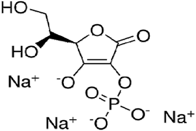
The chemical structure of STAY-C® 50.
2.2. Animals and experimental design
The study protocol was approved by the Institutional Animal Care and Use Committee of Joongbu University (IACUC-20221101-02) and complied with the recommendations in the Guide for the Care and Use of Laboratory Animals. The canines were randomly divided into two groups with three canines in each group: the control group (CG, no treatment) and treatment group (TG, treatment with STAY-C®50). The characteristics of canines used in this study are summarized in Table 1.

Canine characteristics(n=6)
From November 2022 to March 2023, both groups were fed 40 g of commercial feed twice daily. In addition, both the control and treatment groups consisted only of canines that had been diagnosed with stage 1 or higher periodontitis for > 6 months. In the treatment group, sodium ascorbyl phosphate (700 mg) was dissolved in water and applied to the gums near the roots of the teeth using a syringe, in the morning and evening.
2.3. Evaluation of dental plaque
During the experiment, photographs were taken using QScan Plus equipment (AIOBIO, Seoul, Korea) at 0, 2, 4, 6, 10, 12, 14, 16, and 20 weeks to analyze the efficacy of tartar removal using sodium ascorbyl phosphate (Garcez et al., 2020). Photography was performed at consistent illuminance levels and monitored using an illuminometer, while the canine was lying down. The images were transferred to a computer and analyzed using Adobe Photoshop 22 (Adobe Systems Ltd., Europe). We then analyzed and computed the number of pixels for the clear area (white part) and the number of pixels for the colored plaque area (red part) in the incisor plus the canine tooth or the incisor tooth alone. The ratio of the plaque area to the total tooth area was calculated using the following equation (Fig. 2):
- Ratio of plaque area (%) = Number of pixels of plaque area/Number of pixels of total tooth area× 100
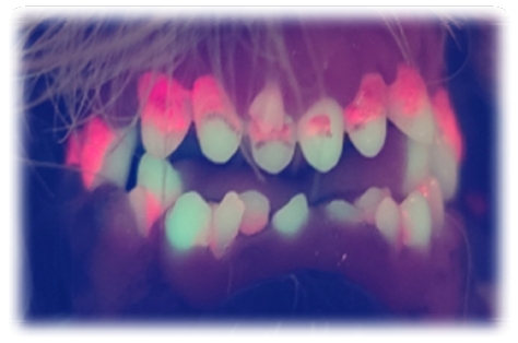
Photograph showing the ratio of the clear area (white part) to the plaque area (red part).
The change in the ratio of the dental plaque area to the total tooth area was determined as the difference between the initial week and the next week’s ratio of plaque area (for example, clear area at 0 weeks–clear area at 2 weeks and clear area at 0 weeks–clear area at 4 weeks). The dental plaque area was calculated in the same manner as the clear dental area.
2.4. Statistical analysis
The results are presented as the mean ± standard error of the mean (SEM). Statistical comparisons between treatments were performed using an independent t-test and carried out using GraphPad Prism 7 software (GraphPad Software, USA). All statistical analyses were performed using SPSS version 12 software (SPSS Inc., USA), and significance was set at p<0.05 for all comparisons.
3. Results and Discussion
Fig. 3 shows the changes in the dental plaque area of the incisors plus canines or incisors alone over time (in weeks). Reduction in the dental plaque area of the incisors plus canines and incisors alone was greater in the treatment group than in the control group over time; however, the differences between the two groups were not significant (p>0.05). To our knowledge, no previous studies have reported the effects of sodium ascorbyl phosphate in companion canines. The most important finding of our study was that the use of sodium ascorbyl phosphate facilitated the enhanced reduction of the dental plaque area of the incisors plus canines or incisors alone. As in humans, dietary texture is an important factor influencing dental plaque, and calculus formation in dogs. Soft foods promote the formation of dental plaque and calculus owing to the lack of abrasive forces during mastication (Gorrel, 1998; Marx et al., 2016). Daily use of a toothbrush in canines is considered the most effective tool for the suppression of dental calculus formation (Hennet, 2006). However, the correct toothbrush and technique must be used, or the gingival tissue may be damaged and the canine will hate the process (Marx et al., 2016). Our results suggest that the efficacy of sodium ascorbyl phosphate may be associated with its combination with iron, which is related to reduced plaque bacterial growth, and the complexation of calcium, which helps reduce tartar formation (Murererehe et al., 2022). Another reason for the high rate of dental plaque in canines is the pH of their saliva. Unlike humans, dogs naturally have more alkaline saliva with pH values ranging from 8.50-8.65 that create a more favorable environment for dental plaque and calculus deposition (Harvey, 2005; Fejerskov and Kidd, 2008; Lavy et al., 2012). In addition, sufficient saliva is required, regardless of the two points above. This implies that salivary calcium and phosphate concentrations or salivary alkaline phosphatase levels should be maintained to balance demineralization and remineralization (Shahrabi et al., 2008). Additionally, vitamin C may be associated with erosive tooth wear owing to its acidity (pH 2.5) (Murererehe et al., 2022). In this study, other possible mechanisms of reducing the dental plaque area could have played a role in maintaining an appropriate pH using sodium ascorbyl phosphate, as it exerts several beneficial effects on the oral health of canines.
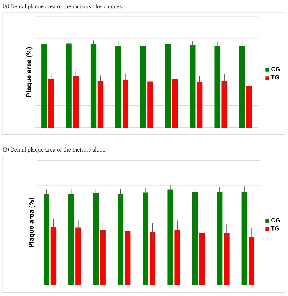
Changes in dental plaque area of (A) incisors plus canines and (B) incisors alone over time (in weeks). Treatments mean: CG = control group (n = 3); TG = treatment group (n = 3). The results are presented as the mean ± standard error of the mean (SEM).
The changes in the ratios of the clear dental area (white part) to the plaque area (red part) in the incisors over time are presented in Fig. 4. For the changes in the ratios of the clear dental area (A) to the plaque area (B) of the incisors, no significant difference between the groups was observed at 0–6 weeks, while significant differences were observed after 10 weeks (p<0.05). Over time, the changes in the clear dental area were significantly greater in the treatment group than in the control group. In addition, the treatment group (that was supplemented with sodium ascorbyl phosphate) exhibited a significant reduction in the dental plaque area (the red part). As shown in Fig. 4, treatment with sodium ascorbyl phosphate appeared to have a short-term effect on the removal of dental plaquecaries in canines, but showed even better results when administered long-term. This suggests that after 10 weeks, the pattern between the treatment and control groups differed, and this difference could indicate a positive response to dental plaque management. The increase in the clear dental area and the decrease in the dental plaque area of the incisors could be explained by the efficacy of sodium ascorbyl phosphate mentioned above.
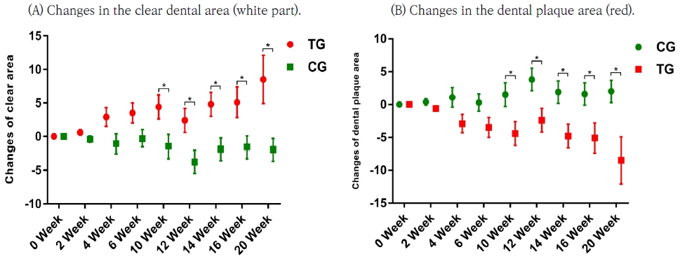
Changes in the ratios of the (A) dental clear area (white part from Figure 2) and (B) dental plaque area (red part from Figure 2) in incisors over time (in weeks). Treatments mean: CG = control group (n = 3); TG = treatment group (n = 3). The results are presented as the mean ± standard error of the mean (SEM). *Bars are significantly different at p<0.05.
4. Conclusions
This study aimed to evaluate the efficacy of vitamin C derivatives such as sodium ascorbyl phosphate in removing dental caries in canines through a preliminary clinical trials. The present study suggests that sodium ascorbyl phosphate could be used to enhance the reduction of the dental plaque area of the incisors plus canines or incisors alone in canines. In addition, treatment with sodium ascorbyl phosphate increased the clear dental area of the incisors and reduced the dental plaque area of the incisors more efficiently after 10 weeks.
REFERENCES
-
Allen, K., 2003, Are pets a healthy pleasure? The influence of pets on blood pressure, Curr. Dir. Psychol. Sci., 12(6), 236-239.
[https://doi.org/10.1046/j.0963-7214.2003.01269.x]

-
Asal, M. A., Abdellatif, A. M., Hammouda, H. E., 2021, Clinical and microbiological assessment of carisolv and polymer bur for selective caries removal in primary molars, Int. J. Clin. Pediatr. Dent., 14, 357-363.
[https://doi.org/10.5005/jp-journals-10005-1956]
- Cho, S. E., Kwon, S. Y., Kim, S. Y., Park, S. Y., Son, N. R., Lee, Y. J., Han, S. Y., 2018, Evaluation of canine oral health status owners and awareness about oral care for companion dogs, Korean J. Clinic Den Hyg., 6, 17-27.
-
EFSA, 2013, Panel on additives and products or substances used in animal feed (FEEDAP), scientific opinion on the safety and efficacy of vitamin C (ascorbic acid, sodium ascorbate, calcium ascorbate, ascorbyl palmitate, sodium calcium ascorbyl phosphate and sodium ascorbyl phosphate) as a feed additive for all animal species based on a dossier submitted by DSM Nutritional Products Ltd, EFSA Journal, 11(2), 3104.
[https://doi.org/10.2903/j.efsa.2013.3104]
-
Eydou, Z., Jad, B. N., Elsayed, Z., Ismail, A., Magaogao, M., Hossain, A., 2020, Investigation on the effect of vitamin c on growth & biofilm-forming potential of Streptococcus mutans isolated from patients with dental caries, BMC Microbiol., 20, 231-241.
[https://doi.org/10.1186/s12866-020-01914-4]
- Fejerskov, O., Kidd, E., 2008, Role of saliva, Den. Caries., 2, 189-208.
- Fontana, M., 1994, Vitamin C (ascorbic acid): clinical implications for oral health–a literature review, Compend., 15, 916-8.
-
Garcez, A. S., Barros, L. C., Fernandes, M. R. U., Fujii, D. N., Suzuki, S. S., Nepomuceno, R., 2020, Fluorescence image and microbiological analysis of biofilm retained around healthy and inflamed orthodontic miniscrews, Photodiagnosis Photodyn Ther., 30, 101707.
[https://doi.org/10.1016/j.pdpdt.2020.101707]
-
Gorrel, C., 1998, Periodontal disease and diet in domestic pets, J. Nutri., 128, 2712S-2714S.
[https://doi.org/10.1093/jn/128.12.2712S]
- Harvey, C. E., 1993, Small animal dentistry, in: Emily, P. P. (ed.), 1st Edition, Mosby-Year Books, St. Luis, 1-143.
-
Harvey, C. E., 1998, Periodontal disease in dogs: Etiopathogenesis, prevalence and significance, Vet. Clin. North Am. Small Anim. Pract., 28, 1111-1128.
[https://doi.org/10.1016/S0195-5616(98)50105-2]
-
Harvey, C. E., 2005, Management of periodontal disease: Understanding the options, Vet. Clin. North Am. Small Anim. Pract., 35, 819-836.
[https://doi.org/10.1016/j.cvsm.2005.03.002]
- Hennet, P., 2006, Canine nutrition and oral health, in: Pibot, P., Biourge, V., Elliott, D. A. (eds.), Encyclopedia of Canine Clinical Nutrition, Royal Canin, Italy, 2006, 388406.
- Jin, H. K., 2017, Clinical characteristics of oral diseases on companion dogs in Gwang myeong City, Master Degree, Chungbuk National University.
-
Kim, S. Y., Youn, G. H., 2014, The relationship between pet dog ownership and perception of loneliness: mediation effects of physical health and social support, J. Inst. Soc. Sci., 25, 215-233.
[https://doi.org/10.16881/jss.2014.01.25.1.215]
- Larsen, J., 2010, Oral products and dental disease, Compend. Contin. Educ. Vet., 32, E4.
- Lavy, E., Goldberger, D., Friedman, M., Steinberg, D., 2012, pH values and mineral content of saliva in different breeds of dogs, Isr. J. Vet. Med., 67, 244-248.
-
Marx, F. R., Machado, G. S., Pezzali, J. G., Marcolla, C. S., Kessler, A. M., Ahlstrøm, Ø., Trevizan, L., 2016, Raw beef bones as chewing items to reduce dental calculus in Beagle dogs, Aust. Vet. J., 94, 18-23.
[https://doi.org/10.1111/avj.12394]
-
Murererehe, J., Uwitonze, A. M., Nikuze, P., Patel, J., Razzaque, M. S., 2022, Beneficial effects of vitamin C in maintaining optimal oral health, Front. Nutr., 8, 805-809.
[https://doi.org/10.3389/fnut.2021.805809]
- Oh, Y. S., Park, J. H., 2020, Emotional state of companion dogs by sensory stimuli, J. Korea Content. Assoc., 20, 199-215.
-
Okajima, L. S., Martinez, E. F., Pinheiro, I. F., Silva, A. S. F., Demasi, A. P. D., 2020, Effect of sodium ascorbyl phosphate on osteoblast viability and differentiation, J. Periodontal Res., 55, 660-666.
[https://doi.org/10.1111/jre.12752]
- Pflipsen, M., Zenchenko, Y., 2017, Nutrition for oral health and oral manifestations of poor nutrition and unhealthy habits, Gen. Dent., 65, 36-43.
-
Shahrabi, M. A., Nikfarjam, J. A., Alikhani, A. A., Akhoundi, N. B., Ashtiani, M. C., Seraj, B. A., 2008, A Comparison of salivary calcium, phosphate, and alkaline phosphatase in children with severe, moderate caries, and caries free in Tehran’s kindergartens, J. Indian Soc. Pedod. Prev. Dent., 26, 74-77.
[https://doi.org/10.4103/0970-4388.41621]
- Smith, B., 2012, The ‘pet effect’: Health related aspects of companion animal ownership, Aust. Fam. Physician., 41, 439-42.
Bio Interactions Lab., BRD Corp.sankim@brdc.biz
Division of Pet & Companion Animal Science, Joongbu University animalscience@naver.com
Division of Pet & Companion Animal Science, Joongbu University doctorj1@joongbu.ac.kr
Division of Pet & Companion Animal Science, Joongbu Universitytaehochung@daum.net